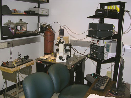
The Nanonics MV 1000 Near Field Scanning Optical Microscope was installed in June of 2006. It is housed in the Molecular and Biological Imaging Center suite of laboratories within the Byrd Biotechnology Building.
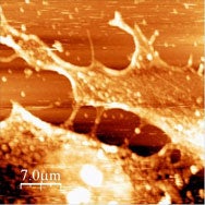
Cultured cells were one of the first samples imaged with this system.
 ....
....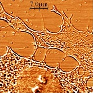
Simultaneously acquired AFM image (left) and transmitted light NSOM image (488 nm) of cultured fibroblast cells on glass cover slip. Note broadening of features in AFM image due to finite fiber tip size.
Near Field Scanning Optical microscopes operate on the principle that an optical aperture or light source (either a hole in a scanning probe or a sharpened fiber optic or an irradiated nanoparticle emitter) when brought into near contact with the sample (distances much less than the wavelength of the light of interest are termed near field), can sense changes in optical properties of the sample at length scales much smaller than the resolution achievable with traditional optical systems.
These tools and others are utilized to aid in caharcterizing and understanding nanomachines, molecular systems and cell biology through research. To discuss use of the system and/or other imaging systems of interest, please contact Dr. Michael L. Norton or refer to the Contact page for further information.
Click for image of NSOM | Lab View 1 | Lab View 2 |
