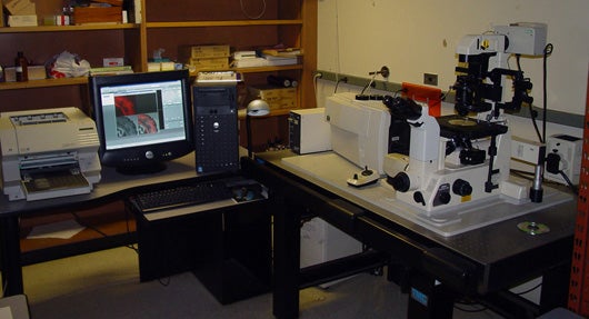


Biorad/Zeiss MRC 1024 with TLD (Transmitted Light Detector) on Inverted Nikon Diaphot 300 with DIC Optics and 6.2 Megapixel Nikon SLR with Mecury Lamp Epifluorescence.
The confocal microscope is a form of microscopy that permits examination of living cells without leading to their demise. Using an argon laser light source, confocal microscopy produces sharp fluorescent images because light from only one focal plane is used to produce the image. Light from other "out of focus" planes is blocked by the detection pinhole, part of the optical system of the microscope (see an animation of how the confocal microscope works). The emitted/reflected light passing through the detector pinhole is distributed into three different wavelength channels, transformed into electrical signals by photomultiplier tubes and displayed on a computer monitor screen.
There are a number of benefits associated with the use of the confocal microscope. These include:
• Light rays from outside of the focal plane will not be recorded.
• Defocusing does not create blurring.
• Scanning the object in x/y-direction as well as in z-direction allows true, three-dimensional data sets of voxels (volume elements) to be recorded.
• Software analysis enables viewing the object from all sides.
• Due to the small dimension of the illuminating light spot in the focal plane, high resolution is obtained.
• By image processing, many slices can be superimposed (called image stacking), giving an extended focus image which can only be achieved in conventional microscopy by reduction of the aperture and thus sacrificing resolution.
•With this microscope resolution is as low as 200 nanometers in XY and 350 nanometers in z axis.
Related links:
• http://www.olympusconfocal.com/java/confocalsimulator/index.html
An interactive simulation made by Olympus of a similar confocal microscope.
• http://rsb.info.nih.gov/ij/
NIH Imagej free download for image analysis.
•http://www.nephrology.iupui.edu/imaging/software/confocal-assistant.exe
Confocal Assistant free download for simple image analysis.
These tools and others are utilized to aid in characterizing and understanding nanomachines, molecular systems and cell biology through research. To discuss use of the system and/or other imaging systems of interest, please contact Dr. Michael L. Norton or refer to the Contact page for further information.
Click for image of Biorad/Ziess MRC 1024| Nikon Diaphot 300 | Lab View 1 | Lab View 2 | Lab View 3

