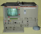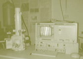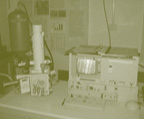


The Scanning Electron Microscope (SEM) is the premiere instrument in the generation of topographical images due to its unbelievable resolution, depth of field and ease of use. Resolution is the ability to discern one point from another, and the wavelength of rays is the factor limiting resolution in microscopy. With a typical light microscope, visible light which has wavelengths between 4,000 to 7,000 Angstroms, the resolution is limited to about 1000 diameters magnification. In contrast, the SEM uses high energy electrons with a wavelength of 0.12 Angstroms, lending to this instrument a limit of resolution of 1 million diameters magnification.
The SEM's principle component is a focused beam of electrons passing through a vacuum and making contact with a dry solid of interest. Those electrons bombard the sample and release secondary electrons. The SEM's primary imaging method is through the collection of those secondary electrons that are ejected from the sample. These electrons are transduced by a scintillation material, which is a phosphor that produces flashes of light when excited by these electrons. The light flashes are then detected and amplified by a photomultiplier tube.
The SEM does more than allow for excellent micrographs to be generated. The X-ray analyzer matches an element to its signature peaks, leading to its proper identification. This analyzer has a beryllium filter on it, meaning that anything smaller than Na will not be effectively detected or identified. The x-ray analyzer is of great use when looking at an imperfection at the elemental level. If the imperfect sample of interest is a manufactured product, this could be the first step in finding the problem. This could then be followed by sampling the assembly line and determining exactly where the imperfection was introduced.
The instruction booklet informs the scientist about the proper use of Marshall's scanning electron microscope (saved as a pdf document - available soon).
These tools and others are utilized to aid in understanding nanomachines, nanotools and related elements through research in nanotechnology. Nanochemistry is an important component of our success and is being investigated by Dr. Michael L. Norton. Refer to Contact page for further information.
Click for image of Oxford EDX | JEOL 5310-LV | Lab View 1 | Lab View 2 | Lab View 3

