
M.U. SEM facility
..
 |
M.U. SEM facility
|
.. |
| .
|
|
 |
 |
|||||||
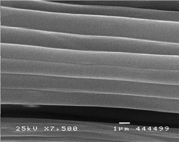
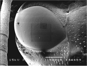
Spider silk (left) and wasp eye (right) demonstration images from unknown species. Note the mild electron beam damage (squares) on the wasp eye. |
|||||||||
|
|
|
 |
||||||
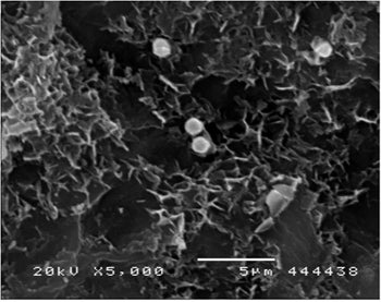
| At left is a strawberry leaf with stoma visible. At right is an arabidopsis leaf; small gold beads are apparent. These were coated with nucleic acids and fired from a ‘gene gun’ in an attempt to locally transform the transcriptional activity in the plant. | |||||||||
 |
|||||||||
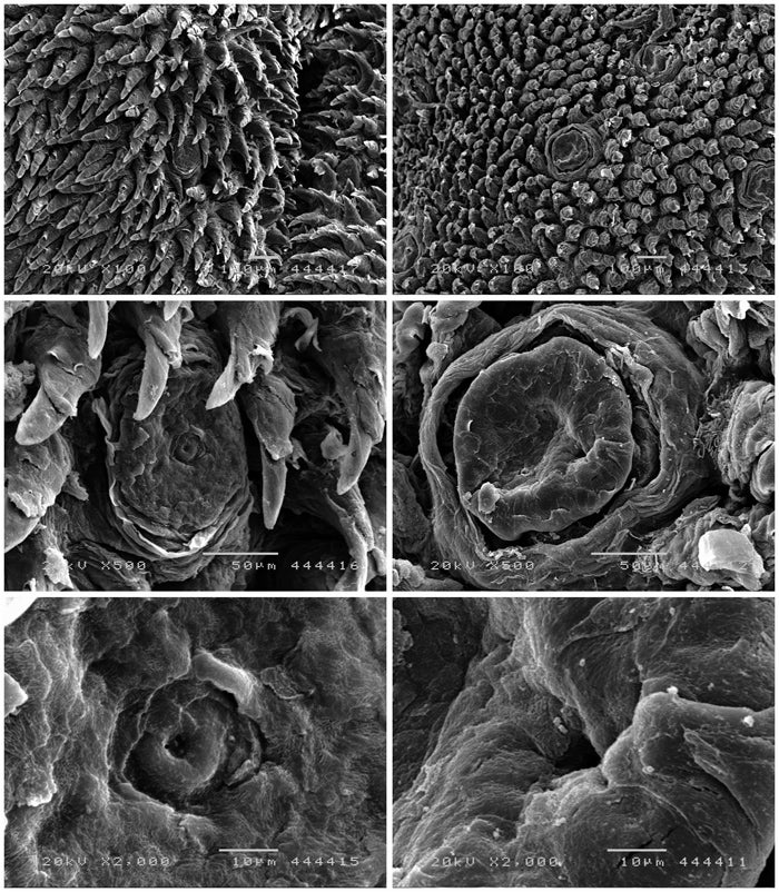
The left column are the fungiform papilla of a rabbit tongue, the right column are those from a rat tongue |
|||||||||

|
 |
||||||||
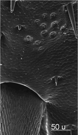
| At right is an sem image of the proximal tibia of P.americana(cockroach) showing the campaniform sensilla. Above is a high mag view of a sensilla (Zill lab). | |||||||||
 |
 |
||||||||
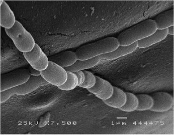
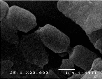
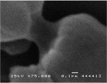
Hydrogen producing cyanobacteria from the lab of Dr. Markov |
|||||||||
 |
 |
||||||||
. |
Biofilm organisms grown in a bio-reactor or collected on glass substrates placed in local streams (Somerville lab). | 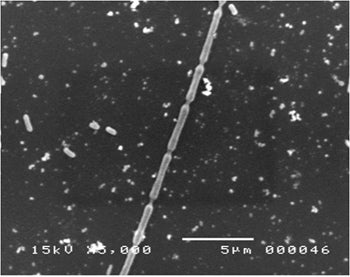 |
|||||||
|
|||||||||