
M.U. Confocal and AFM Facilities
.
 |
||||
M.U. Confocal and AFM Facilities |
||||
. |
||||
| .
|
|
 |
||||||||
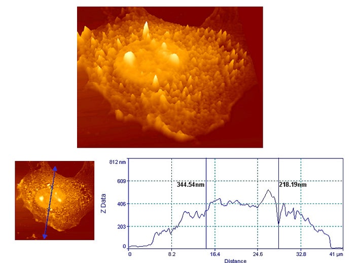
| Topographic representations of a7r5 cells as seen with the MU afm. At top is colchicine treated cell (flattened nuclear perimeter). At bottom is an untreated cell in which a raised peri-nuclear ring is visible. Colchicine disrupts the microtubular sleeve that is believed to surround the nucleas. | |||||||||
|
|
|
|||||||
. |
|||||||||
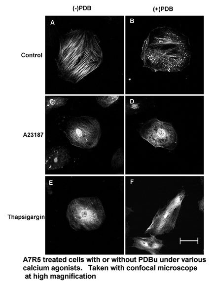
| Confocal images showing microtubular (β-tubulin), microfilamentous (α-actin,phalloidin), and intermediate filamentous (vimentin) cytoskeletal structures in smooth muscle cells (A7r5). Experimental group was treated with phorbol dibutyrate. | 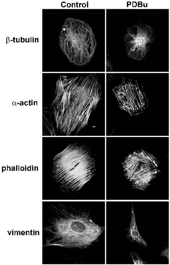 |
||||||||
 |
|||||||||
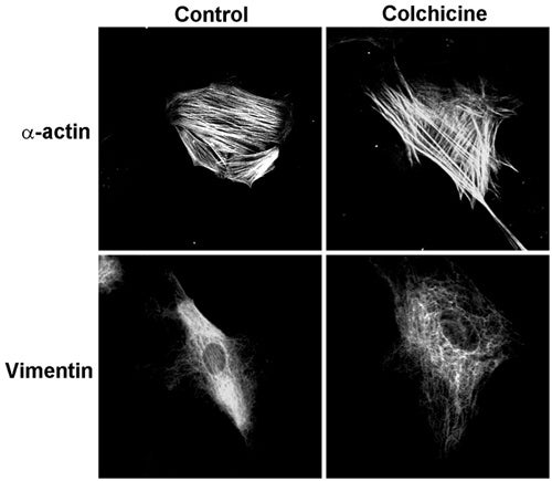
Cytoskeletal effects of colchicine additions to the same smooth muscle cell types. |
|||||||||
 |
|||||||||
 |
|||||||||
 |
|||||||||
 |
|||||||||
|
|||||||||
|
|||||||||