M.U. Confocal Facility
 |
||||
M.U. Confocal Facility |
||||
| .
|
|
 |
 |
|||||||
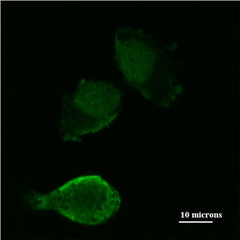
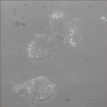
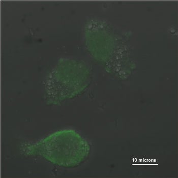
| These cells were human melanoma cells from the radial growth phase stage (early stage melanoma). They were transiently transfected with pLenti-D-TOPO-HIF-1a-785 (lentiviral vector expressing hypoxia inducible factor 1 alpha variant 785). They were then fixed and stained with fluorescine-labled V5 epitope antibody overnight. The HIF-1a785 was tagged with the V5 epitope which allowed us to recognize this fragment of the expressed protein with this florescent labeled V5 antibody. The expected results were that the majority of the HIF-1a785 would localize to the nucleus of these cells as it is a transcription factor. It seems from the picture that the majority of the HIF-1a785 is actually in the nucleus. | |||||||||
|
|
|
 |
||||||
| Montage of optical sections, each is 400nm distant from the last (in z-axis.) X/Y dimensions are 70um x 10um. | |||||||||
|
|||||||||